
Blood flows through a closed system. Blood always travels through blood vessels except when it enters the chambers of the heart. When blood is pumped from the heart, it travels through five types of blood vessels – arteries, arterioles, capillaries, venules, and finally veins.
Arteries are the first type of blood vessels that blood enters after being pumped out of the heart, and when the heart generates pressure and pushes the blood into the arteries, the vessels need to expand to accommodate all of the blood. Therefore, arteries are strong, elastic blood vessels that are designed to carry blood under very high pressure. The strength and elasticity of arteries is made possible by their three-layered walls. These three layers are: the Tunica Interna (inner layer), the Tunica Media (middle layer), and the Tunica Externa (outer layer).
The Tunica Interna is composed of 2 types of tissue: endothelium, which is simple squamous epithelial tissue, and loose connective tissue. The tissues of the Tunica Interna secrete clotting inhibitors that prevent platelets from sticking together when flowing in the bloodstream. Since the Tunica Interna is the inner layer of an artery, it comes in direct contact with the blood, so clotting inhibitors are secreted directly into the bloodstream.
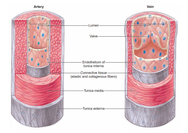
The Tunica Media is the largest and thickest layer of the artery, and it is made of smooth muscle. The smooth muscle in the wall of the artery allows for vasoconstriction, the narrowing of the lumen, or vasodilation, the widening of lumen. Muscles have the functioning property of contracting, so when the muscles in a blood vessel contract, it makes the blood vessel smaller. It is incredibly important to always remember that smooth muscle is INVOLUNTARILY controlled as opposed to skeletal muscle which we can control voluntarily. Additionally, the muscle of the Tunica Media allows the artery to maintain its form and rigidity against the high pressure blood flowing through it. The muscle actually allows arteries to put its own pressure on the blood to keep the blood flow under control and prevent the bursting of a vessel. The Tunica Externa is the outer layer, and it is made up of loose connective tissue, which allows for the elastic property of arteries.
Even though the heart is one unit, it can be thought of as two isolate pumps. The right heart receives venous blood, deoxygenated blood, from the entire body and then pumps that blood to the lungs through the pulmonary artery, a short and quickly branching blood vessel. Like all arteries, the pulmonary artery carries blood away from the heart, but unlike other arteries, it transports deoxygenated blood. Furthermore, the blood pressure in this artery is significantly lower than that of other arteries of the body. The reason is that blood flowing in the lungs meet less resistance than blood flowing through the tissues and organs in the rest of the body. So, because it’s easier for blood to flow through the lungs, the right ventricle is less muscular and contracts with less force than the left ventricle, even though they both eject the same volume of blood. This lesser contraction results in the lower pressure in the pulmonary artery. Again, the idea of form follows function and vice versa is very present in the human body.
After the blood is oxygenated in the lungs, it is transported back to the heart by the pulmonary vein. Similarly to the pulmonary artery, the pulmonary vein differs from the rest of the venous blood vessels of the body. The pulmonary vein, like all other veins, travels towards the heart, but it transports oxygenated blood instead of deoxygenated blood. The oxygenated blood from the pulmonary vein is dropped off in the left atrium and then travels down to the left ventricle after atrial contraction. During systole, the left ventricle pumps the oxygenated blood into the aorta, the largest artery in the body, under a great head of pressure and distends its walls. In diastole, the aortic walls will spring back to its original position due to the composition of arterial walls. Arterial blood is of the fastest speed and highest pressure, which requires arteries to have the greatest amount of elasticity. The aorta branches out into smaller arteries that transport blood to the rest of the body. These smaller arteries, carrying oxygenated blood, continue to divide into smaller branches known as arterioles. The term arterioles literally translates to ‘little arteries’. Arterioles are similar in structure to arteries, but are just smaller in size. As arterioles decrease in size, they lose the tunica externa and tunica media layers. As a result, they will just consist of the endothelium of the tunica interna and bands of individual smooth muscle cells. The arterioles get smaller and smaller until they branch out into capillaries. A ring of smooth muscle, known as the precapillary sphincter, regulates the blood flow from arterioles into capillaries. (Term: Sphincter refers to a ring of smooth muscle).

Capillaries are the smallest blood vessel in the body, and they are composed of just endothelium, which is simple squamous epithelium. The lumen of the capillary measures approximately 0.01 millimeters, allowing for blood to be transported to every single cell in the body. It is important to note that blood travels the slowest at the capillary level. Slow moving blood in a thin blood vessel allows for the efficient diffusion of substances from the blood to the body’s tissues and cells. Think about it, if blood was racing through a capillary, there would not be enough opportunities for the oxygen and nutrients to leave the bloodstream and diffuse into the cells and tissues. Maximum diffusion of nutrients and oxygen is only possible because of the slow moving blood.
But why does blood travel the slowest in our capillaries? The reason is that the blood is so far away from the heart by the time it reaches the capillary networks. The heart is the source of the power for blood flow, so the farther and farther the blood gets away from the heart, the less pumping force actually reaches the blood to propel it forward in the blood vessels. The heart’s force is practically non-existent by the time the blood reaches the capillaries. Another reason for slow blood flow is friction. Friction occurs as the blood passes through smaller and smaller vessels which squeeze the blood and allow less blood to flow through at a time. This ultimately causes the blood flow to slow down.

Capillaries go in between, around, and inside organs to supply them with nutrients and oxygenated blood, and they end up forming large networks of vessels known as capillary beds. If more oxygenated blood is needed for the cells, the precapillary sphincter, which regulates blood flow into the capillaries, would relax, therefore allowing more blood to enter the capillaries. This directly results in more diffusion of oxygen. The contraction of the precapillary sphincter would occur if less oxygenated blood was needed in the capillaries. After giving up oxygen and nutrients while also taking in carbon dioxide and waste products from cellular metabolism, the blood flows from the capillaries into small veins known as venules that later merge together to form veins.
Veins have the same three layers as an artery. However, the difference between the structure of a vein and the structure of an artery lies in the thickness of the Tunica Media, the middle layer of smooth muscle. The Tunica Media of a vein is much thinner than that of an artery. The lack of smooth muscle in a vein lets it have a very flexible shape that can change in accordance with the amount of blood flowing through it. The shape and composition of a vein is completely different to those of an artery. Furthermore, the lack of smooth muscle in the walls of a vein results in a larger lumen, which is the hollow passageway that blood flows through. A larger lumen gives the vein the ability to hold more blood. It is very important to remember that all the veins in the body, except for the pulmonary vein, transport deoxygenated blood, blood with carbon dioxide, back to the heart.
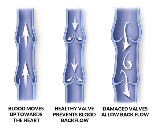
Throughout the body, the pathway of veins parallels that of the arteries. When thinking about the structure of the body in anatomical position, the majority of our organs and tissues are located below the heart. Furthermore, arteries deliver oxygenated blood to all of the body’s tissues, so when most of the body’s tissues are located below the heart, the majority of arterial branches will be heading downwards in our body. This means that blood in the arteries is flowing in the direction of the gravitational pull. On the other hand, veins have to deliver blood back up to the heart, so the flow of venous blood opposes the gravitational pull. When blood tries to move up through veins, there is always the possibility of it flowing back down, resulting in blood pools and not enough blood being delivered to the heart. To combat the potential backflow of blood, veins utilize valves. One-way valves encourage venous blood flow by preventing the backflow of blood. But what is truly the force that pushes venous blood up the veins against the force of gravity? Unfortunately, the thin layer of smooth muscle in the veins is not enough to forcefully contract and push the blood in it upwards. This means that there needs to be another factor that plays a role in encouraging venous blood flow. This factor is the musculoskeletal system.
The role of the musculoskeletal system is dependent on the placement of blood vessels in the body. Arteries are deep in the body, while veins are more superficial. Since veins are superficial, the contraction of skeletal muscles helps to push venous blood up to the heart, especially venous blood in the appendages, where there is more skeletal muscle. However, there are veins located deep in our trunk, but there is not enough skeletal muscle in the trunk to pump the venous blood. This is where the respiratory pump plays a role. When you inhale, your lungs and rib cage expand and increase in size. This larger lung and rib cage actually push up against the veins and squeeze them, which also helps to pump venous blood up to the heart. It is important to note that the respiratory pump only works when you inhale, because only during inhalation do your lungs increase in size due to the intake of air. The veins in the body transport the deoxygenated blood to the right side of the heart via the superior and inferior vena cava. From the right side of the heart, the cycle starts once again, with the pulmonary artery directing deoxygenated blood to the lungs for it to become oxygenated.

The interconnectivity of the cardiovascular system is clearly seen through its network of integrated blood vessels. The function of these blood vessels, the transportation of blood and diffusion of gasses and substances to the tissues, is made possible by their intricate yet simple structures. The cardiovascular system plays such a huge role in the survival of the human body, and the failure of one part in the entire system can and will negatively affect the body. In our next blog, we will cover the circulatory dynamics at play and take a look at common cardiovascular conditions.

One thought on “Blood Vessels”