
Blood needs to leave the ventricles under a certain amount of pressure and at a certain amount for it to properly circulate in the body. The pressure that the blood travels under is dependent on the contraction force of the ventricles as well as the resistance that the head of the blood meets in the arteries and following blood vessels. The main resistance is the inertia of the blood that is already in the blood vessels throughout the body, because the stagnated blood serves as a barrier to the blood flowing through the arteries.
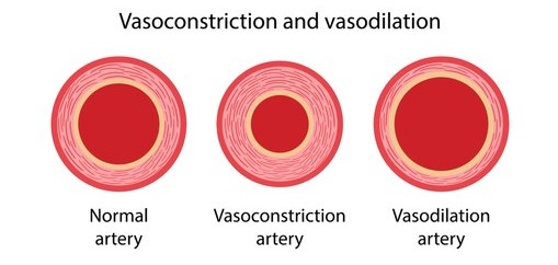
The arterioles, small and thin arteries, are the main regulators of resistance in the body. When the rings of smooth muscle, the tunica media (muscular) layer of the arterioles, contract, the lumen of the arterioles decreases in size, therefore, the pressure and resistance to the blood increases. On the other hand, if the arteriole relaxes, the lumen of the arteriole increases in size, resulting in less resistance, so blood flows more easily and is under less pressure.

The arterioles also directly maintain pressure on the blood during diastole. If the arterioles were completely relaxed during systole, all of the blood entering the arteriole system would rush into the tissue capillaries. To prevent this from happening, the arterioles contract and prevent a fraction of blood from rushing into the capillary system. The blood that is held back will help to exert pressure on the blood vessels during diastole, because more blood in a blood vessel means greater blood pressure. Furthermore, if adequate diastolic pressure is to be maintained in the body, the aortic valve must be able to tightly close. The normal limits of blood pressure are 70 to 90 milliliters diastolic and 110 to 140 milliliters systolic. At these normal blood pressures, the heart and the rest of the body, basically all of the body’s tissues, are supplied with the adequate amount of blood from the arteries needed for them to continue to survive and function. A common trend seen in the general population is that the blood pressure of an individual tends to rise as a person ages. So aging is correlated to the rise in blood pressure.
It is crucial to keep in mind that an individual’s blood pressure will change over the course of a day, and will also vary on a day to day basis. Emotional stimuli, such as sudden fear and anger, leads to an increase in blood pressure because the artery and arteriole walls constrict due to nervous system function and the release of hormones, specifically adrenaline and norepinephrine. Vasoconstriction, the constriction of blood vessels, results in a higher blood pressure because the blood is squeezed by the tightened blood vessel walls. When an individual experiences happiness, their blood vessel walls relax, which is known as vasodilation. Vasodilation results in the lumen of the blood vessels expanding in size, therefore leading to a lower blood pressure. For most people, changes in blood pressure due to emotional stimuli are only temporary, and an individual’s blood pressure will fluctuate according to their emotions and other stimulus factors.
As previously discussed in the Blood Vessels Blog, the nervous system controls the flow of blood through arterioles. The nervous system has the ability to signal a group of arterioles supplying blood to a certain part of the body to constrict more than those of another group of arterioles supplying blood to a different part of the body. The constriction of arterioles is made possible by the rings of smooth muscle on the arterioles and the precapillary sphincters, which are rings of smooth muscle that control how much blood enters a capillary. This mechanism allows the body to control where blood needs to go in the body, so if a certain part of the body is in need of more oxygen (found in hemoglobin in red blood cells), the brain lowers the blood flow to a part of the body that does not need as much oxygen through the constriction of the arterioles and precapillary sphincters, which keeps extra blood available for parts of the body in need of oxygen. For example, when an individual undergoes surgery, they lose a great amount of blood, so the arterioles in the kidneys and intestines contract to lower blood flow to these organs. This makes more blood available for the heart and brain, which are the organs that are needed for the body to continue to survive, especially after a surgery.

When blood enters the capillaries, it experiences a significant drop in pressure and velocity, which is explained by simple mechanics. Arterioles branch out into smaller tubules, which are the capillaries, so it makes sense that the pressure and velocity would drop when blood is split and directed into smaller tubules as opposed to all the blood travelling through one large tubule. Capillary branches interconnect with one another, forming capillary beds around organs, resulting in a further drop in blood pressure and velocity. The low blood pressure and low blood speed in capillaries, in addition to the thin capillary walls allow for substances in the blood, such as oxygen and nutrients to easily diffuse from the blood into the surrounding tissue. The pressure of the tissues as well as the path of the capillaries forces the tissues to push waste products of cellular metabolism, most importantly carbon dioxide, into the bloodstream. It is easier to understand the previously discussed concept as an exchange between the blood and the tissues. The blood gives oxygen and nutrients to the tissues, and in return, the tissues give carbon dioxide and waste products to the blood.

Now the branched capillaries carry deoxygenated blood, blood with carbon dioxide, and they begin to merge together to form venules. These venules then merge together to form veins. Veins are located superficially, so when the muscles in our limbs contract, the veins are squeezed. The squeezing of veins results in the veins being “milked”, and the blood in the veins is pushed up towards the heart. When the limb muscles relax, the deoxygenated blood is prevented from flowing back down the vein because of the closed venular valves. It is important to remember that a series of valves are located along the course of a vein. The contraction of muscles in the limbs opens the valves and pushes the blood forwards through the vein, but when the muscles relax, the valves close and serve as a barrier that prevents the backflow of blood.
However, if the valves in a vein break down and are unable to close completely, the blood will flow backwards and will pool at the lowest working valve. This causes the vein to distend and stretch out, which is what leads to varicose veins. Varicose veins are enlarged veins that have pools of blood in them. The enlarged varicose veins tend to push through the skin to become very visible to the eyes, and they are usually found in the legs. A common side effect of varicose veins is anemia because not enough blood is reaching the heart due to a good amount of it pooling in the veins. When varicose veins occur in venules in a small and concentrated area, the condition is known as Telangiectasia, more commonly known as spider veins. Varicose veins and spider veins are usually treated with compression stockings to squeeze your legs from the outside to push the blood up towards the heart. Other common treatments include exercise or surgery to close off those veins or remove them. The surgery option is usually suggested when the varicose veins are becoming a problem to an individual’s daily life or interfering with their health.


Deep Vein Thrombosis (DVT) is another common condition that affects the veins. DVT is basically a blood clot that develops in veins deep in the body and it mostly affects veins in the lower limbs, like varicose veins. DVT occurs to people who sit down for long periods of time because the skeletal muscles in the legs are in a relaxed state as opposed to being in a state of contraction, like when someone is walking. As covered before, the contraction of skeletal muscle in the legs pushes venous blood up to the heart, but relaxation of skeletal muscle results in blood sitting in one place for a long time. So, when blood is stagnated in the body for a period of time, the nutrients leave the blood and stick to the vein walls, forming a clot. This clot is known as a thrombus, and if the thrombus breaks free and travels with the venous blood, it will enter the heart and get pumped out with blood into the aorta, which branches out into smaller arteries. Arteries get smaller and smaller, unlike veins which get bigger and bigger. So, when a thrombus travels through an artery, it can get stuck in the vessel, and essentially cut off blood flow to areas of the body that the blocked vessel feeds. Without sufficient oxygen being delivered to the tissues of the body through the blood, those tissues will start to die. Deep vein thrombosis is most commonly treated with anticoagulants, also known as blood thinners. Blood thinners help to combat the formation of blood clots, which removes the main risks of DVT caused by a blood clot blockage. Other treatments of DVT are thrombolytics, which are drugs that help to break up clots and compression stockings.
Interesting Facts About Veins:

- Sixty to seventy percent of all blood in the body is travelling through veins and venules at any given time. The reason for this is because blood is travelling at a very slow speed and under very little pressure in veins when compared to the high blood pressure and high blood velocity in arteries.
- When you go to the hospital to get your blood drawn for donation or tests, the nurse puts the syringe into your vein for two reasons.
- 1) Most veins are superficial, so they are much easier to reach than arteries, which are located deep inside the body.
- 2) The pressure in veins is less than that of an artery, so there is less bleeding when the syringe is taken out of a vein. The blood pressure in arteries is very high, so if you took the syringe out of an artery, blood would spurt out everywhere and it would be harder to heal the damage in arteries. Veins are the complete opposite of arteries because they have low blood pressure and low blood velocity, which is what makes it easier to draw blood from veins.
In our next Anatomy and Physiology blog post, we will cover the basic functions of the lymphatic system as well as its connection to the blood and the cardiovascular system.
