
The cardiovascular system is the system of blood circulation. The term cardiovascular, can be broken down into “cardio”, which means pertaining to the heart and “vascular”, which refers to the vessels of our body that contain fluid, specifically blood. Therefore, the cardiovascular system involves the heart, blood, the arteries, the veins, and the capillaries of the human body.
It is crucial to understand that all of the cells within our body rely on the cardiovascular system to provide them with vital substances, specifically oxygen and the nutrients from the food we consume, and remove waste products, such as carbon dioxide, from them. The circulatory system is the system that circulates blood around our body which keeps our whole body functioning. For example, the circulatory system transports hormones released into the bloodstream to cells in the body through the networks of arteries and capillaries. Though the circulatory system is what keeps our body functioning, it is monitored by nervous and endocrine systems, so, damage to other organs such as the pancreas, lungs, or brain will affect the circulatory system and its function.
There are actually two circulatory systems, usually known as the “greater” circulatory system and the “lesser” circulatory system. The lesser circulatory system consists of the heart, the lungs, and their interconnecting blood vessels in the body, and is known as the pulmonary circuit. The greater circulatory system is comprised of the heart and all of the vessels that supply the remainder of the body with blood, and is known as the systemic circuit. There are very distinctive differences between these two systems, but in both systems, the heart is the primary factor.
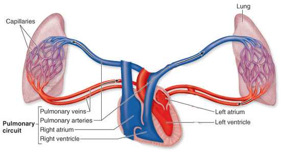
Although the heart is considered one unit both functionally and anatomically, it can actually be thought of as two separate pumps, the “pulmonary heart” which is the right side of the heart, and the “systemic heart” which is the left side of the heart. The only route of communication between these two pumps is the lungs. The journey of blood throughout the body starts in the right side of heart. So the right heart receives deoxygenated blood (blood with carbon dioxide) from the veins in the body and pumps it into the lungs by the way of the lesser circulatory system. In the lungs, the blood is oxygenated (supplied with oxygen) and is then moved to the left side of heart. From the left heart, the oxygenated blood is pumped into a large artery called the aorta, which distributes the blood to the entire body through the greater circulatory system.
Essentially, the heart is a hollow organ composed mostly of muscle that is divided into four chambers. The right heart is made up of an upper chamber called the atrium or auricle, and a lower chamber called the ventricle. Between these two chambers is a one-way valve that is called the tricuspid valve. The left heart has its own left atrium and ventricle, but the valve that separates its upper and lower chamber is called the mitral valve. So, both the right and left heart have their own atrium, ventricle, and a valve that allows blood to pass from the upper chamber to the lower chamber.
The heart pumps blood to the rest of the body by “beating”. By beating, I mean an unceasing rhythmic contraction and relaxation, a feature and function that is unique to the heart. The “beating” phenomenon can be explained in the following way. The heart is composed of two distinct cells. The majority of these cells are similar to the muscle cells that are attached to our bones to allow voluntary movement, except, the heart muscle fibers(cells) are interconnected with one another. The other type of heart cells are found in isolated areas, and these cells are specialized structures that are crucial to transmitting nervous impulses to the heart. Collectively, all of these specialized cells are known as the Purkinje system, named after the scientist who discovered them.
The heart is able to beat because of the electrical impulses transmitted from the cardiac conduction system. The impulse that causes the contraction of the heart to begin is found in one of the isolated areas of specialized heart muscle, called the sino-atrial node. For clarification, a node is a mass of specialized fibers. This impulse is an electrical impulse that triggers our heart to contract, and therefore pump blood. So, from the sino-atrial node, the impulse spreads over to the left and right atria. This impulse causes both atria to contract simultaneously, which is known as atrial syncytium. There is no specialized transmitting tissue, like in the sino-atrial node, in the atria, but the interconnecting muscle fibers of the heart relay the impulse to one another. Another node called the atrioventricular node is found where the atria meet the ventricles. The atrioventricular node receives the impulse from the atria, and after a short pause, it directs the impulse to the major segment of the Purkinje system.
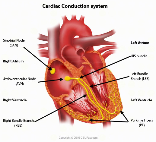
The major segment of the Purkinje system is called the Bundle of His, which is located between the left and right ventricles. The Bundle of His branches out into the left and right branches which reach the apex of the heart and spread out across both ventricles. These branches allow both ventricles to receive the impulse simultaneously. Like with the atrial muscles, the impulse is quickly transmitted to all the fibers of both ventricles. The impulse spreads throughout the ventricles and causes ventricular syncytium, which is the simultaneous contraction of the left and right ventricle.
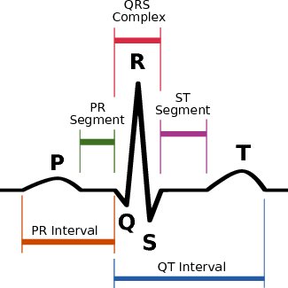
The impulse spreading throughout the heart can be recorded by an electrocardiogram, which produces a graphic tracing of the electric current. The time it takes for the electrical impulse to travel from the sino-atrial node to the entire heart is only a mere fraction of a second. After the impulse spreads throughout the heart, only then does the actual contraction of the atria and ventricles occur in succession.
It is common knowledge that the heart has a basic rhythmic pattern, but the speeding up and slowing down of the heart rate, the pulse, is actually controlled by the nervous and endocrine system. For example, the injection of adrenalin, also known as epinephrine, increases the heart rate. On the other hand, the stimulation of the vagus nerve results in the heart rate slowing down. As the amount of blood entering the right atrium increases, then the heart rate speeds up. Our heart rate is also accelerated by an overactive thyroid gland or an excessive administration of the hormone thyroxine.
The heart is definitely a more efficient pump than any man-made gadget. The heart’s function is to pump out blood to the rest of our body, which it can only do when it receives blood through blood vessels, specifically the veins. Blood from the rest of the body, returns to the heart through two large veins known as the venous trunks. These two venous trunks are called the inferior vena cava and the superior vena cava. The inferior vena cava transports blood from the lower parts of the body, specifically the lower extremities, abdomen, and parts of the chest, hence the name “inferior” vena cava. The superior vena cava transports blood to the heart from the upper parts of the body, specifically the upper extremities and the head. Both of these venous trunks empty into the right atrium, which is where the deoxygenated blood is delivered to the heart.
The atria, both the left and right, have very thin-walled chambers when compared to the ventricles because the main job of the atria is to let blood flow down into the ventricles. The atria have the ability to easily adapt to incoming quantities of blood because they have attached auricles. Auricles serve as an extra storage chamber for blood when there is excess blood being delivered to the atria. It is very important to understand that blood pours into the atria when the heart muscles are relaxed. Now, when the heart is relaxed, the atrioventricular valves, the valves that are situated between the atria and ventricles on both sides of the heart, are open. This allows blood to continuously flow into the relaxed ventricles. The state of relaxation of the heart is known as diastole.
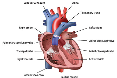
An impulse sent from the sino-atrial node travels to the muscles of the atria and causes the atria to contract. When the atria contracts, the chamber size decreases and the pressure inside the atria increases, which causes the blood in the atria to be pushed out into the ventricle. To be able to reach the ventricle, the blood needs to pass a valve which opens when the pressure in the atria increases. The valve on the right side of the heart is called the tricuspid valve and the valve on the left side is called the bicuspid or mitral valve.
The wave of contraction that started in the atria, travels down the ventricles. When the ventricles contract, the chamber size decreases and the ventricular pressure increases. When the pressure inside the right ventricle increases, a column of blood under great pressure is forced up and out, which causes the tricuspid valve to tightly close and opens the pulmonary valve. The pulmonary and aortic valves are the valves that separate the ventricles and the blood vessels, respectively the pulmonary trunk and the aorta. When the pulmonary valve opens, the blood enters the pulmonary arteries which takes the deoxygenated blood to the lungs. On the left side of the heart when the same process occurs, the pressure generated inside the left ventricle causes the mitral valve to close and opens the aortic valve, which allows the oxygenated blood to flow into the aorta, which branches out into various arteries across the body. The period of contraction of the atria and the ventricles is called systole.
As diastole comes again to the heart chambers, the blood that was pushed into the pulmonary arteries and the aorta, tries to return back to the ventricles due to the suctional backflow pull that ventricular diastole causes. This backflow of blood is prevented by the closing of the pulmonary and aortic valves. This cycle of diastole and systole occurs approximately 65 to 90 times a minute.
For the heart to function properly, it’s valves need to be working. The four valves of the heart, the mitral, tricuspid, aortic, and pulmonic valve are not composed of muscle tissue, instead they are composed of tough collagenous tissue. The mitral valve, located between the left atrium and ventricle, looks like a parachute that is cut in half. The two flaps of the parachute, also known as leaflets, are attached to chordae tendineae, which are strands of fibrous connective tissue. The chordae tendineae are anchored to papillary muscles located on the inner wall of the left ventricle. The chordae tendineae are attached to the valve flaps in such a way that allows the two flaps to tightly close and shut the passageway between the left atrium and ventricle.
Let’s compare the function of a parachute to the function of a valve. A parachute is inflated by an inrush of a column of air. With the mitral valve, it is an inrush of ventricular blood that causes the valve flaps to move together and close the passageway between the left atria and ventricle. When the pressure in the ventricle increases and the ventricular blood shoots up, the mitral valve flaps swing back up, meet as a horizontal line, and tightly fit together, with the chordae tendineae keeping them in place. The tricuspid valve, the other atrioventricular valve, separates the right atrium and ventricle and functions the same way the mitral valve does. The only difference, however, is that the tricuspid valve has three flaps instead of two, hence the name “tri-cuspid”.
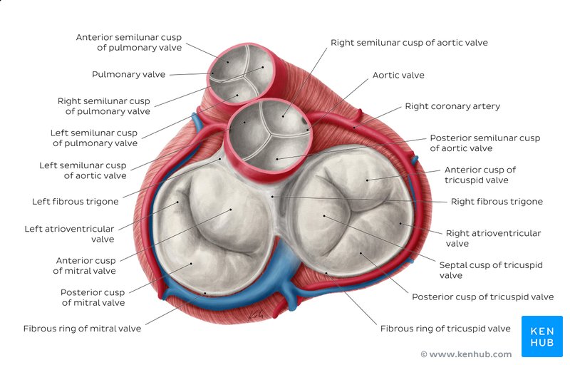
The aortic and pulmonic valves, collectively known as the semilunar valves, are built differently from the atrioventricular valves, the mitral and tricuspid valves. The semilunar valves are structured like hinges. For example, the aortic valve is situated where the aorta and left ventricle meet. The aortic valve is constructed of three cusps, which project out of the walls of the aorta. When the left ventricle contracts, the pressure inside increases as the size of the ventricle becomes smaller. Ventricular systole pushes up the ventricle blood till it strikes the cusps of the aortic valve causing the cusps to open outward, which allows the ventricular blood to freely flow into the aorta. But when the ventricles undergo diastole, the ventricles expand, therefore causing the pressure inside the ventricles drop. Ventricular diastole results in the backward pressure of blood in the aorta which causes the aortic valve cusps to swing shut. When the aortic valves close during the relaxation period of the ventricles, it prevents the blood that entered the aorta from flowing back into the ventricle. The pulmonic valve functions in the same way that the aortic valve does but is located between the right ventricle and pulmonary trunk.
When the valve cusps are disrupted in their shape or if something keeps them from opening and closing properly, then the valves can no longer function properly which will result in the disruption of blood flow. The incomplete closure of a heart valve is known as a prolapse. A prolapse can lead to blood, oxygenated or deoxygenated, flowing in the wrong direction, and it results in an audible backflow of blood. A heart murmur is the noise heard because of a prolapsed valve.
Like all the other muscles and tissue in the body, the heart muscles are supplied with oxygenated blood by the left and right coronary arteries. The coronary arteries are the first arteries that branch off of the aorta. So, during ventricular diastole, the coronary arteries fill up with oxygenated blood to carry to the heart. On the other hand, the rest of the arteries in our body fill up with blood when the ventricles undergo systole.
There are two main reasons why the coronary arteries fill up with blood during diastole. When the heart is in systole, the cardiac muscle contracts, which narrows the branches of the coronary arteries that are embedded within the heart. Furthermore, in systole the blood pushed into the aorta is travelling away from the heart under great speed and pressure, so only small amounts of blood would be able to enter the coronary vessels. However, the backflow pressure present in diastole forces sufficient quantities of blood into the coronary vessels.
Overall, the heart plays the main role in the function of the cardiovascular system. Because the cardiovascular system is so complex, in this post, I have only covered the anatomy of this system. In the next blog post, I plan to write about the circulation aspect of this amazing system.
