
The extent of human knowledge has greatly increased over the recent years, with inquiring individuals making breakthrough discoveries in many fields. Despite their achievements and intellectual abilities, most people lack an understanding of their bodies, the structures within, as well as the collaboration between those various parts.
As I take you on the journey to understand the “normal” human body, I will first cover the meaning of “normal” in regards to anatomy. For those who are unaware of its definition, anatomy is the study of structures in the human body. On the other hand, physiology, which is often paired with anatomy, is the study of the functions of the structures in the human body at different complexities. The parts of the body are not formed by automated machines that are programmed to create our organs the same way. Instead, our bodies are developed through many combatant forces such as hereditary factors and environmental influences, which come together to create the human body. It is extremely important to understand that no two individuals are exactly the same. The concept of a “normal” human being is a compounded picture of what is considered the most common biological findings.
Facts and ideas change all the time in biology, as biologists and scientists challenge the known knowledge with new discoveries. Before we get into discussing the body and its various functions, I need to clarify some of the terminology used to reference our anatomy. The key living unit of the body is the cell, and a variety of different cell types are found in every part of our body. Additionally, several cells with similar behavior and functions assembled in a group are called a tissue.
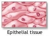
Epithelial tissues are found throughout the body. Epithelial cells form epithelial tissues by being arranged in groups without any fibers in between each cell so that they form compact lining membranes. Layers of the epithelium, also known as epithelial tissue, cover the body’s surface and makeup part of the skin. There are various types and shapes that epithelial tissue comes in, each with its specific function. For example, glandular epithelium forms secretions and lines the stomach and intestine. On the other hand, most of the liver is formed by epithelial tissue arranged in cords.
Other examples of specialized tissue include
- Muscle tissue
- Osseous/bony tissue
- Nervous tissue
- Adipose/fatty tissue
In addition to these specialized tissues, there are also less commonly known types of tissue which include
- The endothelium, which lines the body’s vascular channels, such as blood vessels
- The mesothelium, which covers the internal body cavities such as the abdominal cavities or the chest
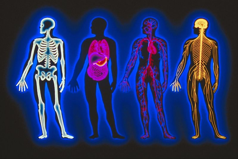
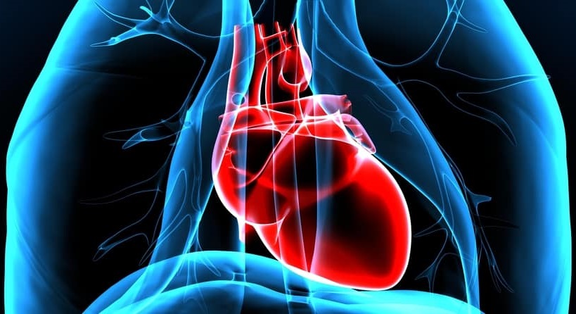
It is important to remember that when 2 or more types of tissues are grouped together in a specialized pattern, they form a distinct structure called an organ, which performs a specific function. Examples of an organ include the heart, the skin, and the lungs. Furthermore, a group of organs and structures that are interconnected and work together to perform one common specialized function is called a system.
The human body, in several ways, can be compared to a country. A country is composed of many states, which in the human body represent the organs. In these states, we have the ultimate unit, the individual, which equals a cell in the human body. However, this analogy fails when we acknowledge the behavior and functions of organs as a system. For example, the respiratory system is a group of organs from the nose to the lungs that collaborate with one another to let air, specifically oxygen, enter into the body and to eliminate gases, like carbon dioxide, from the lungs. The reproductive system includes the organs and passages that are involved in the formation and development of a child.
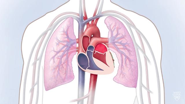
These organ systems cannot function independently. For example, the respiratory and reproductive systems both require the activity of all the other systems to be able to function properly. The respiratory system cannot continue to function without proper blood supply which is provided by the circulatory system. Similarly, the heart, part of the circulatory system, would stop beating without sufficient oxygen from the lungs.
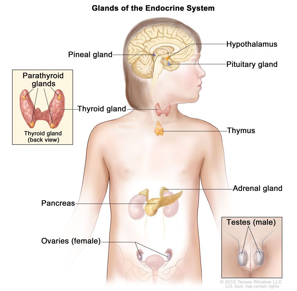
A system does not need to be localized to a specific part of the body. Most of a system’s components are located in different parts of the body, away from one another. For example, let’s look at the endocrine system. The pituitary gland is located in the brain and is far away from the other glands of the endocrine system, such as the thymus which is located in the lungs and the pancreas which is located in the stomach. Another example is bone marrow. Bone marrow is part of the lymphatic system, not the skeletal system, yet bone marrow is encased by the skeleton.
As I mentioned before, the smallest living unit in the body is the cell. A body cell is a structure that is enclosed by a delicate membrane that limits the fluid and materials that flow in and out of the cell. The cell’s membrane is crucial to the function of the cell, and ultimately the survival of an individual.
A cell is composed of many different parts. Within the cell, the nucleus is the largest as well as the most essential component, which has its own semi-permeable membrane. Inside the nucleus lies the nuclear genes. Nuclear genes are unique hereditary factors that shape the individual that bears them and are passed onto the future cells that the individual will also come to bear. These genes are arranged in thread-like material called chromatin, which comes together and pairs up to form chromosomes. The remaining materials of the cell, such as the mitochondria and the vacuole, make up the cytoplasm. The cytoplasm contains various structures and substances, however not all these can be observed using a regular microscope. By using techniques that utilize a high-speed centrifuge to isolate particles by their weight, and an electron microscope to provide high magnification, we are able to recognize structures within the cytoplasm.
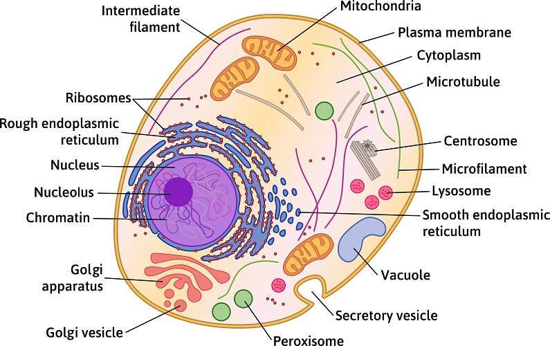
Some of these structures contain enzymes, which are a group of chemical substances that catalyze and accelerate numerous chemical reactions and transformations in the body. A few structures are protein-based, while other structures represent granules of secretion due to the specialized activity of cells.
The cells of different tissues and organs vary in appearance, making them easily identifiable. The cells vary in shape, size, contour, number of nuclei, cytoplasmic material, and function. A way to be able to recognize these different cells is by their appearance in a group and a tissue, along with their relationship between one another, leading to the formation of glands, cords, or interlacing fibers.
Cells are usually cultivated in the body, however, they can be cultured in media apart from the body, and when properly cared for, can reproduce amongst themselves. In cultures other than the human body, many of the cells’ unique characteristics persist, however, the pattern and period of growth are individually based amongst cells.
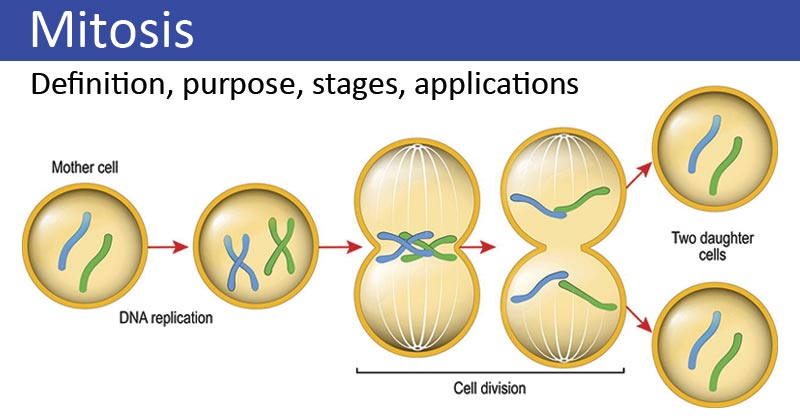
Through the human body’s existence throughout an individual’s life, cells go through the cell cycle, meaning that old cells die and are replaced by new cells. Cell division is accomplished through the process of mitosis. During mitosis, the chromatin material located in the cell’s nucleus separates into chromosomes, which become aligned vertically in the middle of the nucleus. The nuclear membrane fully disappears and each chromosome is split down the middle into 2 halves. When the two sets of chromosomes, the halves, have moved apart, a nuclear membrane forms around each set. The cytoplasm then divides in half, thereby creating 2 independent and identical cells.
Cell division in the human embryo during development is not restricted to the reproduction of one kind of cell. If cell division only led to the creation of just one type of cell, we would never have different organs and tissues, each with their own specific functions. It is crucial to understand that the reproduction of cells is kind of an unequal division where some daughter cells become “differentiated”. There are two categories of differentiation concerning cells. A highly differentiated cell has specialized characteristics in appearance and function, but has lost the ability to grow and mature into a variety of cell types. On the other hand, a poorly differentiated cell can also develop into various cell types, but it lacks the functional abilities of a specific differentiated cell.
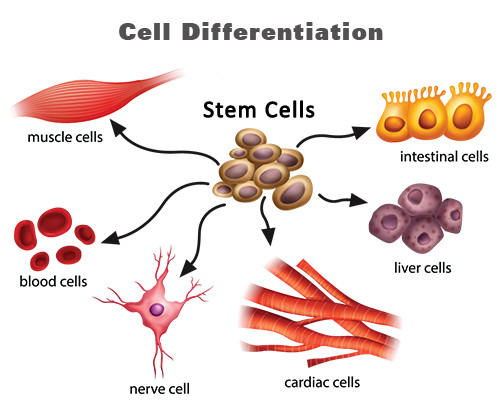
Both cell types are usually present in our organs, however, an exception would be our brain. In the brain, nerve cells are in development during the embryonic stage, but as the human body grows old, the nerve cells begin to degenerate. The degenerated nerve cells in our body are not replaced during our lifetime. Another exception is the skeletal muscles and the heart muscles, which are limited to very slight growth in regards to reproduction. They can still grow and increase in size by taking in additional materials into the cells that compose the muscle, however, the number of cells will not increase.
In all organs and tissues, there is a greater amount of functional cells than is necessary for the adequate performance of the organs and tissues. Additionally, the body continues to maintain this superfluous supply of cells. For example, if a segment of the liver is damaged or even removed, the remaining healthy cells will rapidly reproduce to make up for the lost/damaged area. Another example would be if one kidney were taken out of the body, the other kidney would grow in size to compensate for the loss of the first kidney. However, the loss of nerve cells is a permanent deprivation, only compensated if other nerve cells assume the role and take on the function of the cells destroyed.
As the organs develop in the human embryo, a process other than multiplication and differentiation occurs. This process is known as organization, and it pertains to the formation of bodily structures in a concrete pattern. Organization occurs due to the growth of specialized cells as well as the fibers of connective tissue to act as the supporting framework. For example, if kidney cells are cultivated through micropropagation, the growth of tissues or cells in a medium outside the body, they grow in thin sheets and do not form tubules like they would if they grew in the body. However, if connective tissue cells are added, the kidney cells growing outside the body can organize and form structures.
The importance and function of connective tissue can be recognized from its name. Connective tissue is found throughout the body, forming valves, ligaments, and tendons in all organs and tissues that surround the individually functioning structures, like the heart or liver. Three different types of fibers comprise the category of connective tissue. The finest fibers are called reticulin fibers, which form an intricate network throughout all the organs that work as the main supporting structure in the body. Collagenous fibers are composed of collagen. The thickness of these fibers allow it to provide a strong framework for the body and form the capsules that surround the organs. Finally, elastic fibers are formed by elastin and are located primarily in tissues that are frequently subjugated to expansion and contraction. Elastic fibers are found in the lungs, skin, and even the walls of blood vessels.
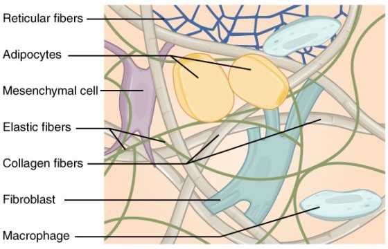
Researchers have figured out that these fibers are connected to connective tissue cells called fibroblasts and fibrocytes. Researchers still don’t exactly know how fiber is formed, however, they have observed that connective tissue fibers form readily in a substantial quantity when an injury leaving a gap in the tissue has occurred. These fibers are also the part of scar tissue that can grow in any part of the body.
Along with the connective tissue fibers in our body, there is a mucus-like fluid called the ground substance, which fills in the tissue spaces throughout the body. The characteristics of the ground substance vary based on different locations in the body and at different times according to the passage of various materials from the cells and blood through it.
Previously, connective tissue and ground substance held very little interest to medical researchers. However, in recent decades, connective tissue has been recognized as the location for the alterations that lead to allergic reactions. Nowadays, a lot of attention is being focused on trying to understand the nature, characteristics, and hidden functions of connective tissue. The discovery of hydrocortisone, a secretion of the adrenal gland, has shined a light on the effects of the adrenal gland on the state of connective tissue.
The situation involving the repair and regeneration processes of the tissues and cells of the body is easily carried out. For example, if a couple of functioning cells are lost, new cells will grow and mature in their place. If the framework in an area of tissue is lost, scar tissue will rebuild and patch up the framework. However, if an entire organized structure, like a heart or kidney, is lost, no new organ will grow back in the human body to replace the lost one. The organ structures are too intricately composed by different tissues, that they are unable to be developed or replaced by highly differentiated cells in the body.
Studying human anatomy is a complex process because of the intricacy of the body. By breaking down the human body into smaller, less complex systems, the task of understanding anatomy and physiology becomes much easier. In the upcoming posts of this blog, we will discuss the different organ systems along with the interrelating components of the human body that help an individual function and survive. The cardiovascular system is the topic we will be covering in the next post!
